Ontario Animal Health Network (OAHN) Bovine Expert Network Quarterly Veterinary Report
Interesting Cases from the Animal Health Laboratory Q2 Bovine Data
A total of 1512 bovine cases were submitted to the AHL during Q2, spanning from May 1 to July 31, 2023. Of these, 193 submissions had a pathology component, with 44 postmortem cases and 149 send-in cases (including 73 meat inspection cases). Submissions from practitioners included 61 dairy, 52 beef, 2 bison, and 5 where the commodity was not specified. Insufficient clinical history was associated with 7 submissions.
Case 1: Mycoplasma wenyonii in a Jersey Cow
History: Mature jersey cow, 6 DIM, history of abrupt drop in milk, red urine, decreased appetite, pale mucus membrane.
Findings: On-farm necropsy findings- pale icteric membranes, yellow tinged liver, enlarged gall bladder. Bloodwork- marked regenerative anemia.
Microscopic findings- Acute hepatic necrosis and segmental renal tubular degeneration (presumed hypoxic injury), rare renal intra-tubular pigment (suspect hemoglobin). EDTA blood- PCR detection of Mycoplasma wenyonii, negative for Anaplasma marginale and A. centrale; Kidney- PCR negative for Leptospira spp.
Summary: This cow had marked regenerative hemolytic anemia with PCR detection of Mycoplasma wenyonii in the EDTA blood sample, and infection with this hemotropic parasite combined with the immunosuppression associated with calving/post-partum period was the presumed cause of this animal’s rapid clinical deterioration and death. Extravascular hemolysis would be incited by the presence of the hemotropic parasite. It may be that intravascular hemolysis was occurring as well, as M. wenonyii infection has previously been suggested as a trigger for immune-mediated hemolytic anemia in a cow (1). It appears most cattle infected with M. wenonyii do not develop clinical disease, but clinical illness will arise in animals that become immunosuppressed. Though not described in this case, some additional clinical signs that may be reported in affected animals include swollen teats, edema of the distal hindlimbs, prefemoral lymphadenopathy, transient fever, weight loss, diarrhea, and reproductive inefficiency (1, 2).
- Gladden N, Haining H, Henderson L, Marchesi F, Graham L, McDonald M, Murdoch FR, Bruguera Sala A, Orr J, Ellis K. A case report of Mycoplasma wenyonii associated immune-mediated haemolytic anaemia in a dairy cow. Ir Vet J. 2016 Jan 22;69:1. doi: 10.1186/s13620-016-0061-x. PMID: 26807214; PMCID: PMC4724101.
- Genova SG, Streeter RN, Velguth KE, Snider TA, Kocan KM, Simpson KM. Severe anemia associated with Mycoplasma wenyonii infection in a mature cow. Can Vet J. 2011 Sep;52(9):1018-21. PMID: 22379205; PMCID: PMC3157061.
Case 2: Ductal plate malformation in a poor performing calf
History: Several poor-doing calves over the last few months (5 months of age). Eating and drinking as they should but not growing as well as their pen-mates. No clinical signs of pneumonia or scours, lung sounds normal, no fever.
Findings: Hepatic lesions were compatible with a ductal plate malformation and accompanying pancreatic dysplasia and fibrosis. This calf also had bacterial bronchopneumonia with isolation of high levels of Pasteurella multiocida and Histophilus somni.
Summary: The hepatic lesion was presumed to be congenital in nature, stemming from a lack of proper ductal plate remodelling during development, and an underlying genetic abnormality (either sporadic or inherited) was suspected. The congenital lesion in the liver and pancreas would explain this calf’s poor growth, and bacterial pneumonia may have been contributing to poor performance in this calf as well as its pen-mates.
Case 3: Clostridiodes difficile detected in neonatal calves
History: Two approximately 10-day-old calves found sick and puffing. Treated with antibiotics and electrolytes and died the next day.
Findings: On farm PM identified diarrhea. Microscopic examination of intestine revealed cryptosporidia, mild neutrophilic colitis in 1 calf, and lesions suggestive of sepsis/toxemia.
Summary: These calves had diarrhea and lesions suggestive of septicemia, but the exact cause was not determined. Bacterial culture was performed on one fecal sample (source uncertain), and 3+ Clostridiodes difficile (formerly Clostridium difficile) was isolated, though the exact clinical significance of this finding is unclear. PCR testing was negative for rotavirus and coronavirus, and 1+ cryptosporidium was identified. Bacterial culture of lung was performed for both calves, which yielded moderate levels of E. coli isolated in mixed culture, and PCR testing for Salmonella Dublin was negative on both lung samples. Advanced autolysis hindered microscopic assessment of small intestine, and culture was not performed on intestine from either calf, so the possibility of diarrhea and septicemia associated with E. coli remained. Clostridioides difficile is a gram-positive, spore-forming bacterium that is an important cause of disease in people. It has been identified as a potential contributor in the development of diarrhea in young calves, but further work is needed to investigate this possibility. In a 2006 study (1), C. difficile was detected in dairy calves in Canada, with isolation from 31 of 278 calves, representing 11 of 144 diarrheic calves (7.6%) and 20 of 134 (14.9%) control calves. In a 2008 publication on C. difficile in calves (2), the organism and its toxins were identified in approximately 25% of stool samples from diarrheic calves, and diarrheic calves were found to be almost twice as likely to be culture positive.
- Rodriguez-Palacios A, Stämpfli HR, Duffield T, Peregrine AS, Trotz-Williams LA, Arroyo LG, Brazier JS, Weese JS. Clostridium difficile PCR ribotypes in calves, Canada. Emerg Infect Dis. 2006 Nov;12(11):1730-6.
- Melissa C. Hammitt, Dawn M. Bueschel, M. Kevin Keel, Robert D. Glock, Peder Cuneo, Donald W. DeYoung, Carlos Reggiardo, Hien T. Trinh, J. Glenn Songer. A possible role for Clostridium difficile in the etiology of calf enteritis. Veterinary Microbiology. Volume 127, Issues 3–4. 2008, Pages 343-352.
Clostridium difficile was reclassified as Clostridiodes difficile in 2016 to better reflect differences between C. diff and members of the genus Costridium. The common name C.diff is maintained.
| About This Report |
| This summary has been compiled by Dr. Rebecca Egan, Animal Health Laboratory (AHL) from diagnostic submissions to the AHL Guelph and Kemptville locations. |
Have an idea for an infographic you’d like to see, or a podcast you’d like to hear?
Email oahn@uoguelph.ca to let us know!
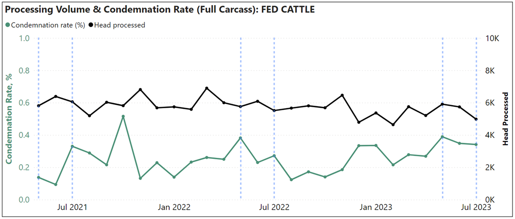
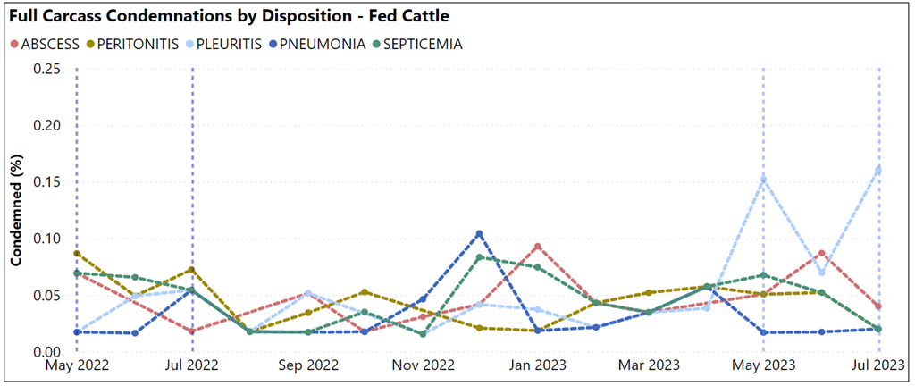
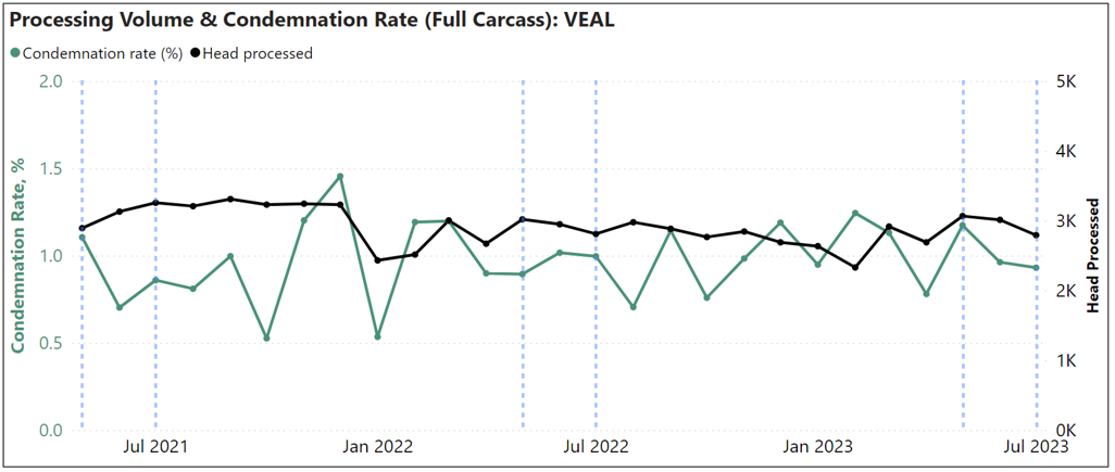
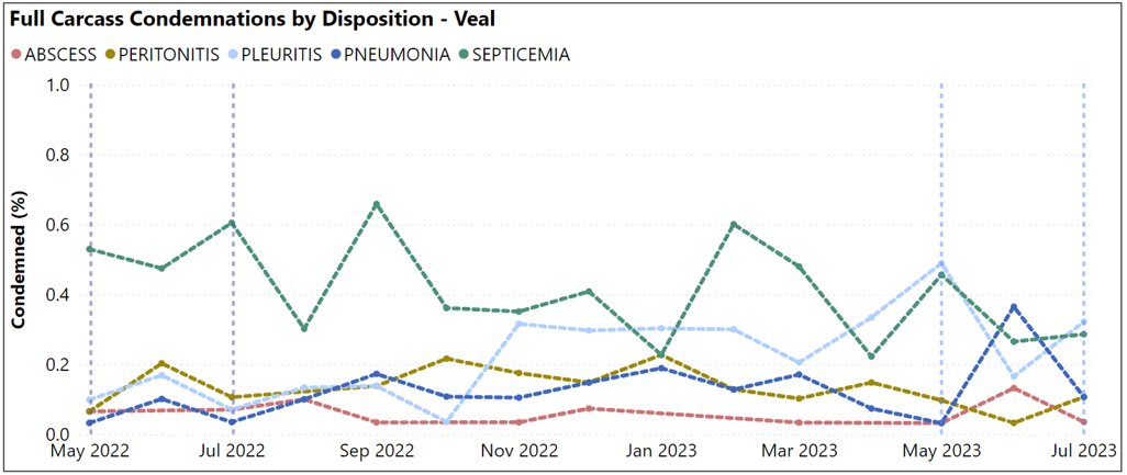
OMAFRA Bovine Provincial Slaughter Condemnation Data
Q2 2023 (May – July)
Fed Cattle:
Veal:
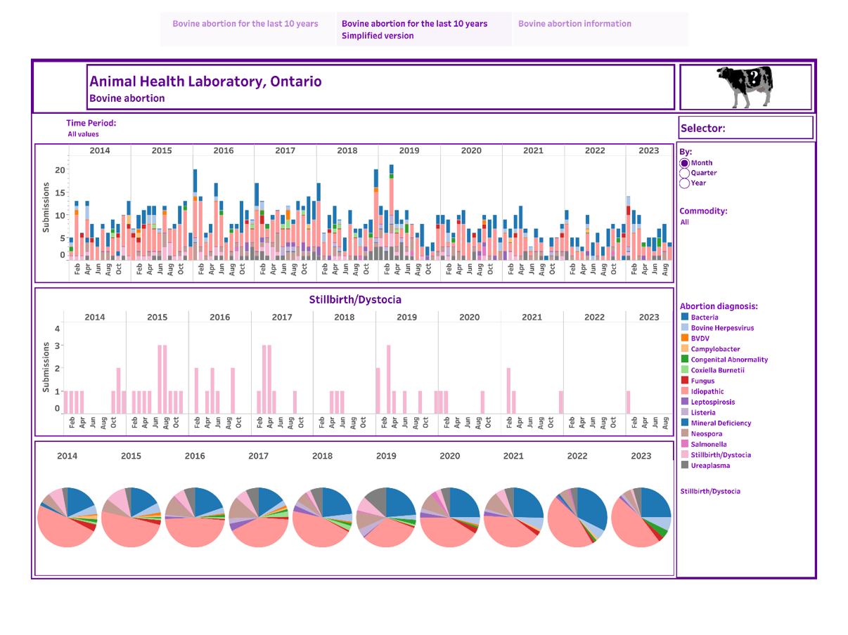
The Interactive Animal Pathogen Dashboard (IAPD) project is pleased to announce the addition of bovine abortion dashboards!
This year the OAHN bovine network provided funding and industry insights for production of bovine abortion dashboards which are now live on the website. These new dashboards display current and historic trends in causes of bovine abortions, allowing practitioners to make more informed vaccine recommendations and track potential emerging or re-emerging diseases. Dashboard interactivity allows users to focus and tailor the visuals to answer specific questions of interest by filtering for time aggregation and commodity group.
Large animal practices and researchers can apply for a free licence by going to our website https://iapd.lsd.uoguelph.ca/ and clicking “Get Access”. Producer groups and commercial entities can purchase licences at cost.
Identification of Culicoides species found in selected areas of Ontario from June – September 2022.
OAHN Project of the Bovine, Equine, and Small Ruminant Networks
Project Lead: Katie Clow with student Valentina Gonzalez Rodriguez
Collaborators: Jocelyn Jansen, Cynthia Miltenburg, Alison Moore, Tanya Rossi
Background
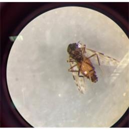
Photo credit: Valentina Gonzalez Rodriguez
Culicoides spp. are a genus of biting midges that are important vectors of several viruses, including bluetongue virus (BTV) and epizootic hemorrhagic disease virus (EHDV) in ruminants. Ontario has many native Culicoides spp. which are not competent vectors of these viruses. However, the recent detection of these viruses in previously unreported areas of Ontario, Canada, suggests the possibility of a northward expansion of new species of Culicoides. BTV serotype 13 was detected in cattle in 2015 and EHDV detected in deer in 2017 and 2021. These changes warrant increased surveillance of these species to identify potential changes in disease risk to wild and domestic ruminant populations. The objective of this project was to describe spatial and temporal patterns of Culicoides spp in select areas of Ontario that have not been previously sampled.
Research Design
In this study, field sampling was conducted at 9 selected sites in Ontario, consisting of ovine, bovine, and equine farms. Two ultraviolet blacklight traps were set up on each premises and run for three consecutive nights every other week from June to September 2022. Trap collections were preserved in a salt solution and then Culicoides spp. were separated from bycatch in the laboratory and stored in 95% ethanol. A subset of samples were morphologically identified using a stereoscope and identification keys. The remaining collections were classified using the environmental DNA (eDNA) in the ethanol preservation solution with high-throughput sequencing targeting the cytochrome oxidase subunit 1 (COI) gene. Spatial analysis was conducted to examine the distribution of 3 different Culicoides species of interest.
Results
A diversity of Culicoides spp. were detected across the farms during the sampling time frame. Culicoides subgenus Avaritia was detected on all premises. This subgenus includes three species that have been confirmed as vectors for BTV in Europe. Further molecular differentiation of a subset of samples confirmed the presence of these three species; C. chiopterus was found on one premises, C. obsoletus on all premises, and C. sanguisuga on four premises. Subgenus Monoculicoides was detected on seven premises. This subgenus includes the known vector species for BTV and EHDV, C. sonorensis. Presence of C. sonorensis was confirmed via molecular methods at 2 premises, one in central Ontario and one in eastern Ontario. Culicoides stellifer is suspected to be a vector for both BTV and EHDV and was detected on all premises.
This study highlights that Culicoides spp. of animal health concern are present in the province in several areas. Given minimal previous surveillance, it is challenging to contextualize these findings. Longitudinal monitoring across areas of southern and eastern Ontario for Culicoides spp. would greatly enhance our ability to monitor changes in the vector population and assess the risk to animal health.
Help us help you!
Have an idea for an infographic you’d like to see, or a podcast you’d like to hear? Email oahn@uoguelph.ca to let us know!


